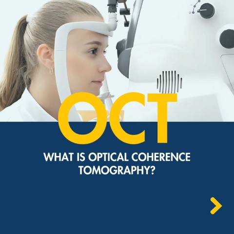

An Optical Coherence Tomography (OCT) test may sound like a mouthful, but the process itself is more straightforward. An OCT is a non-invasive test that allows your optometrist to take detailed cross-section pictures of the back of your eye.
Including an OCT test during your eye exam allows for early detection and monitoring of various eye conditions like macular degeneration, glaucoma, and diabetic retinopathy—often before symptoms present themselves.
Early detection allows for timely intervention and management to preserve your vision.
How Does an OCT Test Work?
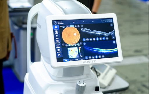
An OCT employs a laser (with no radiation) to obtain high-resolution images of the layers of the eye’s retina and the optic nerve. Your eye doctor can use these colour-coded images to scan for irregularities in the optic nerve, and accurately measure the thickness of the retina.
The clear, 3D images provided by an OCT test can help patients visualize any problems they may be experiencing with their eyesight.
An OCT scan provides a clear map to help your eye doctor find areas of the eye with abnormalities or early damage. It’s vital to catch eye conditions in the early stages to prevent damage to a patient’s vision.
Diagnosing vision problems in the early stages can allow your optometrist to provide preventative treatment for conditions including glaucoma, and retinal diseases like age-related macular degeneration (AMD).
OCT has been described as a safe, non-invasive ultrasound for your eye, using light instead of sound waves to create a map of the retina and optic nerve. Guided waves of light provide a precise 3D image of the eye’s structure, without making direct contact with the patient’s eyes.
Because OCT relies on waves of light, it is not effective for any condition that affects how light passes through the eye, including cataracts.
What Can an OCT Test Detect?
OCT is used to diagnose a variety of eye conditions, like:
- Diabetic retinopathy: This condition occurs when blood glucose levels are high for extended periods, causing damage to your eyes’ blood vessels and the retina.
- Macular holes: This condition occurs when there’s an opening or tear in the macula, resulting in blurry central vision.
- Macular pucker: A macular pucker is a wrinkle in the macula that creates a wavy spot in your central vision.
- Macular edema: When there’s a fluid back-up in your macula, it’s called macular edema. This condition can result in colours appearing washed out, and blurry vision.
- Age-related macular degeneration (AMD): AMD occurs as the macula slowly deteriorates throughout the ageing process.
- Central serous retinopathy: This condition may lead to retinal detachment, which requires emergency care.
- Glaucoma: This condition results from a build-up of fluid at the front of your eye, which can result in damage to the optic nerve.
OCT & Glaucoma
Patients at risk of developing glaucoma can benefit from an OCT scan, as the images can help evaluate the anterior angle of the eye. The anterior angle is an essential part of the eyes’ anatomy that operates as the drainage channel for fluid.
When the drainage channel is not working correctly, it can lead to an increase in eye pressure. This pressure causes damage to the optic nerve and can result in vision loss.
An OCT provides your optometrist with detailed, multi-level images of the anterior angle. These images can help determine severity, and distinguish between types of glaucoma, such as:
- Open-angle glaucoma: Occurs when the eye’s drainage angle remains open, but fluid doesn’t drain properly, resulting in difficulties maintaining standard intraocular pressure.
- Closed-angle glaucoma: Occurs when the eye’s drainage angle closes, which results in a rapid increase in intraocular pressure.
An optometrist can conduct OCT scans in darkened rooms, which allows for quick and accurate results. The position of the eye’s anterior angle in a dark room is a vital step in diagnosing acute closed-angle glaucoma.
Should I Have an OCT Test?
An OCT exam may be recommended by your optometrist as part of your routine eye exam if:
- You are over age 25
- You are at risk of developing eye disease
- You have been diagnosed with an eye disease
- You have any detected abnormalities in your retinal layers
- You are experiencing thickening of retinal layers
- You are being treated for an eye disease that requires monitoring
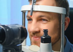
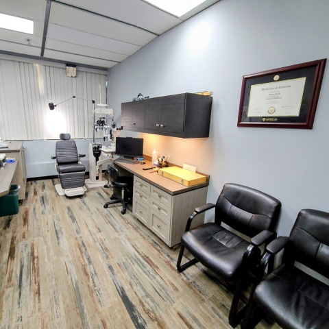
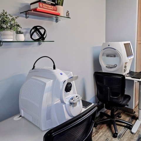
What to Expect During Your OCT Scan
Your optometrist may use eye drops to dilate your pupils, allowing for a more accessible retinal examination. Once your pupils are dilated, you will be asked to sit in front of the OCT machine with your head resting on the support to encourage stillness.
Once in the proper position, your eye doctor will advise you to look into the device’s lens, which contains a blinking target. At this point, the machine scans your eye with no physical contact. The OCT test is completed in approximately 5 to 10 seconds.
If your optometrist used dilating eye drops before the exam, your eyes might be sensitive to light for a few hours afterward.
Keeping a Sharp Eye on Your Health
If you have questions about OCT tests or would like to book an appointment, do not hesitate to reach out to our experienced team at Calgary Family Eye Doctors.